Digital panoramic X-ray for a dental assessment at PureDental
2Dpanoramic X-raya full overview of your teeth
3DCBCT scanbefore implants and oral surgery
Digital diagnostics · Budapest, District II

CBCT and panoramic X-ray

Accurate diagnosis, safer treatment planning

An accurate diagnosis is one of the most important foundations of successful dental treatment. A thorough oral examination reveals a great deal, but some dental problems cannot be seen with the naked eye: hidden inflammation, bone loss, changes at the root tip, the position of wisdom teeth, bone resorption after tooth loss, or the anatomical details needed before implant placement.

In PureDental’s clinic in Budapest’s District II, digital panoramic X-rays and 3D CBCT scans give us a precise picture of the teeth, the jawbone, the maxillary sinus, the nerves and the surrounding structures. This means your treatment plan is based not on guesswork, but on detailed, personalised diagnostics.

Book an appointment for dental diagnostics!
  • Digital panoramic X-ray for a complete dental overview
  • 3D CBCT scan for planning implants, oral surgery and bone grafting
  • More accurate diagnosis of hidden inflammation and bone changes
  • Personalised treatment plan
  • Fast, painless imaging examination
  • Modern digital dental diagnostics in Budapest, District II
2Dpanoramicoverview image of all your teeth
3DCBCTspatial image of bone, nerve canal and sinus
4stepsfrom preparation to evaluation
0injectionspainless, non-invasive examination
2D or 3D?

What is the difference between a panoramic X-ray and CBCT?

Panoramic X-ray · 2D

A panoramic X-ray is a two-dimensional overview image of the mouth that shows the teeth, the jaws, the area around the jaw joints and part of the maxillary sinus at the same time. It is often the first imaging examination before a dental check-up, tooth extraction, wisdom tooth assessment, gum disease evaluation or general treatment planning.

CBCT · 3D

CBCT, or dental CT, on the other hand provides a three-dimensional image of the teeth and jaws. It is especially useful when precise spatial information is needed: for example when planning a dental implant, before bone grafting, for a sinus lift, for complicated wisdom tooth removal, when examining inflammation at the root tip, or before more complex oral surgery.

Put simply: a panoramic X-ray gives a good overview, while CBCT provides detailed 3D diagnostic information whenever it is needed to plan treatment safely.

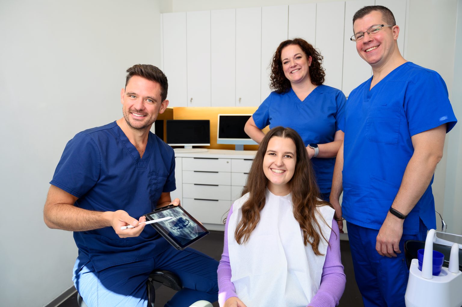
Reviewing a panoramic X-ray with our patient at PureDental’s District II clinic
2D overview image

What is a panoramic X-ray?

A panoramic X-ray is a digital, two-dimensional dental X-ray that shows, in a single image, all the teeth, the upper and lower jaws, the area around the tooth roots, missing teeth, the position of the wisdom teeth and certain bone changes.

One of the greatest advantages of a panoramic X-ray is that it is fast, painless and gives a comprehensive picture of the condition of the mouth. It helps the dentist spot problems that are not clearly visible during an oral examination.

A panoramic X-ray may be taken, for example:
  • as part of a first dental assessment,
  • before drawing up a larger treatment plan,
  • before a tooth extraction or oral surgery,
  • to check the position of the wisdom teeth,
  • to review gum disease and bone loss,
  • to check teeth that had root canal treatment in the past,
  • to assess missing teeth and tooth replacement options,
  • as part of a focal infection assessment,
  • for diagnostics before orthodontic treatment.
First step

A panoramic X-ray is therefore primarily an overview examination. It helps decide which direction further examination should take and whether more detailed, targeted imaging, such as a CBCT scan, is needed.

Evaluating a dental CBCT scan at the PureDental clinic in Budapest
3D imaging

What is CBCT, or dental CT?

CBCT stands for cone beam computed tomography, a three-dimensional dental imaging examination. A CBCT scan provides a detailed spatial image of the teeth, the jawbone, bone thickness, the position of the nerve canal, the floor of the maxillary sinus, the course of the roots and the surrounding anatomical structures.

Dental CBCT is particularly important when a conventional two-dimensional X-ray does not provide enough information for a safe decision. The 3D image allows more accurate planning of the implant position, the extent of bone grafting, the need for a sinus lift, the risks of wisdom tooth removal or the background of a more complex root canal problem.

A CBCT scan may be taken, for example:
  • before dental implant placement,
  • to plan bone grafting or a sinus lift,
  • before complicated wisdom tooth removal,
  • to examine how close a lower wisdom tooth is to the nerve canal,
  • when hidden root tip inflammation or a focal infection is suspected,
  • before failed or repeated root canal treatment,
  • when root resorption, a crack or an anatomical variation is suspected,
  • to map cysts, bone changes and oral surgery cases more precisely,
  • when planning full mouth rehabilitation or complex tooth replacement.
Only when indicated

CBCT is not always necessary, but when it is indicated, it can make the treatment plan significantly more accurate and safer.

A personalised decision

Panoramic X-ray or CBCT: which one is needed when?

Panoramic X-rays and CBCT are not competitors, but imaging examinations suited to different diagnostic situations. We always choose the right image based on the question we need to answer.

First dental assessment

2DPanoramic X-ray – Usually sufficient

Yes, often taken first

3DCBCT – May be needed if…

In case of complex changes, implant planning or complaints of unclear origin

Dental implant

2DPanoramic X-ray – Usually sufficient

Can be useful as an initial overview

3DCBCT – May be needed if…

To plan the implant’s position, angle, available bone and anatomical risks

Bone grafting

2DPanoramic X-ray – Usually sufficient

Can provide an overview

3DCBCT – May be needed if…

To measure bone height, bone width and the defect precisely

Sinus lift

2DPanoramic X-ray – Usually sufficient

Suitable for orientation

3DCBCT – May be needed if…

To examine the sinus floor, bone height and anatomical relationships precisely

Wisdom tooth

2DPanoramic X-ray – Usually sufficient

Often sufficient as a first examination

3DCBCT – May be needed if…

If the roots are close to the nerve canal or the tooth is in a complicated position

Tooth extraction, oral surgery

2DPanoramic X-ray – Usually sufficient

May be sufficient in simpler cases

3DCBCT – May be needed if…

In case of complex root shape, proximity to a nerve, or a suspected cyst or bone change

Root canal treatment

2DPanoramic X-ray – Usually sufficient

Can provide an overview, but a targeted intraoral X-ray is often also needed

3DCBCT – May be needed if…

In case of a hidden canal, root fracture, resorption, retreatment or unclear inflammation

Periodontology

2DPanoramic X-ray – Usually sufficient

Useful for an overview of bone loss

3DCBCT – May be needed if…

For a more precise analysis of complex, localised bone defects or bone loss around implants

Orthodontics

2DPanoramic X-ray – Usually sufficient

Can be part of the initial assessment

3DCBCT – May be needed if…

In case of an impacted tooth, abnormal tooth position or a complex anatomical question

Focal infection assessment

2DPanoramic X-ray – Usually sufficient

Useful as a first step

3DCBCT – May be needed if…

If the nature or extent of the change cannot be determined precisely from the panoramic X-ray

The goal

The goal is not to take the highest-resolution or most detailed image in every case, but to obtain enough information for an accurate diagnosis and treatment plan with the lowest radiation dose necessary.

Indications

When may a CBCT scan be needed?

Before dental implant placement

When planning a dental implant, CBCT is one of the most important diagnostic tools. It allows a precise assessment of the quantity, thickness and height of the available bone, as well as the position of the nerves, the maxillary sinus and the surrounding anatomical structures.

CBCT matters not only for surgical safety, but also because the implant position must always be planned with the future crown, bridge or full-arch restoration in mind.

Learn more: Dental implants →

Based on this, we can determine:
  • where the implant can be placed safely,
  • what implant length and diameter can be used,
  • whether bone grafting is needed,
  • whether a sinus lift is needed,
  • the ideal angle and position of the implant,
  • what final restoration can be planned on the implant.

Before bone grafting

If a tooth has been missing for a long time, the jawbone can shrink over time. In such cases there may not be enough bone to place an implant. A CBCT scan helps assess whether bone grafting is needed and, if so, to what extent.

The 3D image shows more precisely the extent of the bone defect, the thickness and height of the jawbone, and whether bone grafting can be carried out at the same time as implant placement or should be planned as a separate procedure.

Learn more: Dental bone grafting →

Before a sinus lift

In the back of the upper jaw there is often not enough bone height to place an implant safely, because the maxillary sinus lies close to where the tooth roots used to be. In such cases a sinus lift may be needed.

This is especially important because planning a sinus lift requires great precision.

Learn more: Dental bone grafting →

A CBCT scan helps assess precisely:
  • the position of the sinus floor,
  • the available bone height,
  • the condition of the sinus membrane,
  • any anatomical variations,
  • whether an open or closed sinus lift is possible.

Before wisdom tooth removal

The position of wisdom teeth can often be assessed well on a simple panoramic X-ray. However, there are cases where a CBCT scan allows safer planning.

In such cases the 3D image can help plan the procedure and protect the surrounding structures.

Learn more: Wisdom tooth removal →

CBCT may be indicated, for example, if:
  • the roots of a lower wisdom tooth run close to the nerve canal,
  • the tooth lies deep in the jawbone,
  • the roots are curved or follow an unusual course,
  • a cyst or other change is suspected around the tooth,
  • the surgical risk cannot be assessed precisely from an earlier 2D image.

Before oral surgery

Before a complex extraction, apicoectomy, cyst removal, bone grafting or other oral surgery, CBCT can give a more accurate picture of the spatial relationships between the teeth, roots, bone and anatomical structures.

This is especially important if the procedure is close to the nerve canal or the maxillary sinus, or if the exact extent of a change is not clear from the panoramic X-ray.

Learn more: Oral surgery →

Root canal treatment and examining root-treated teeth

For most root canal problems, a targeted intraoral X-ray or a panoramic X-ray may be sufficient. A CBCT scan may be considered if conventional images do not provide enough information or a more complex problem needs to be clarified.

Learn more: Microscopic root canal treatment →

CBCT can help, for example:
  • to find hidden root canals,
  • to assess root tip inflammation more precisely,
  • to investigate why a previous root canal treatment failed,
  • when root resorption or a crack is suspected,
  • when planning an apicoectomy,
  • during a focal infection assessment, if the 2D image does not give a clear answer.

Gum disease and inflammation around implants

In gum disease, a panoramic X-ray can give a good overview of the extent of bone loss. In some cases, however, CBCT may also be needed, for example to assess localised bone defects, deep pockets, furcation involvement or bone loss around implants more precisely.

In such cases CBCT can help refine the periodontal or peri-implantitis treatment plan.

Learn more: Periodontology →

Before full mouth rehabilitation and complex treatment plans

If treatment involving several teeth, implants, crowns, bridges, orthodontics or full mouth rehabilitation is needed, diagnostics are especially important. In such cases, a panoramic X-ray and, if necessary, CBCT together help make the treatment plan logical, transparent and safe in the long term.

Learn more: Dental assessment and diagnostics →

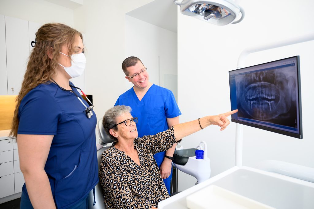
Evaluating a panoramic X-ray with our patient as the first step of treatment
First step

When may a panoramic X-ray be enough?

In many cases a panoramic X-ray is sufficient as a first step, because it gives a comprehensive picture of all the teeth and the jaws. It is particularly useful when the goal is a general assessment, defining the first steps of a treatment plan, or reviewing the wider context of a dental problem.

A panoramic X-ray may be enough, for example:
  • during a first consultation and assessment,
  • to review missing teeth,
  • to check old crowns, bridges and root-treated teeth,
  • to assess general bone loss in gum disease,
  • before a simpler tooth extraction,
  • for a first examination of wisdom teeth,
  • for initial diagnostics before orthodontic treatment,
  • as the first step of a focal infection assessment.
If a more precise image is needed

However, if the panoramic X-ray shows a change that requires more precise spatial examination, or if millimetre-precise information is needed to plan treatment safely, we may recommend a CBCT scan.

The examination

How is a panoramic X-ray or CBCT scan taken?

Both panoramic X-rays and CBCT scans are fast, painless and non-invasive. Before the image is taken, our team member helps you into the correct position, and the device then captures the required image within a few seconds.

1

Preparation

Before the examination, metal objects that could interfere with the image must be removed. These may include glasses, earrings, necklaces, piercings or removable dentures, if required for the given image.

If you are pregnant, may be pregnant, or have had many imaging examinations recently, always let us know before the examination.

2

Positioning

Correct positioning of the head is important for good image quality. You stand or sit next to the device and stay still while the machine rotates around your head. The examination does not hurt, involves no injections and requires no anaesthesia.

3

Taking the image

A panoramic X-ray is usually done in a few seconds. A CBCT scan is also quick, but including positioning it may take a few minutes.

It is important for the patient to stay still during the scan, because movement can reduce image quality and, in extreme cases, the image may need to be retaken.

4

Evaluation

The image is evaluated by the dentist or specialist based on your symptoms, the oral examination and the planned treatment. An X-ray or CBCT image does not replace a personal examination; it complements it.

The goal is to see exactly what is causing the problem, which treatment options may be considered, and which solution would be safe in the long term.

Radiation protection

Safety and radiation exposure

Digital dental X-rays and CBCT involve ionising radiation, so every image must be justified. At PureDental we only recommend a panoramic X-ray or CBCT scan if it is genuinely needed for the diagnosis, the treatment plan or the safe performance of a procedure.

CBCT is an examination of high diagnostic value, but we use it not routinely, only when indicated. This ensures that the scan genuinely adds to the diagnosis and the safety of treatment.

What we take into account

The principle of radiation protection is to obtain the necessary diagnostic information with the lowest reasonably achievable radiation dose. That is why we always consider:

  • 01what clinical question we need to answer,
  • 02whether a smaller or simpler image would be enough,
  • 03whether a usable X-ray or CBCT scan was taken earlier,
  • 04whether 3D imaging is justified,
  • 05how large an area needs to be imaged,
  • 06whether an especially cautious protocol should be used for children or young patients.
What the images show

What does the dentist see on a panoramic X-ray?

A panoramic X-ray shows an overview of all the teeth and the jaws at once. It allows the dentist to examine the condition of the mouth comprehensively and to recognise changes that may be present without any symptoms.

A panoramic X-ray may show:
  • the number and position of the teeth,
  • missing teeth,
  • the position of the wisdom teeth,
  • changes around root-treated teeth,
  • larger areas of decay,
  • bone loss due to gum disease,
  • suspected cysts or other bone changes,
  • asymmetry or abnormalities of the jaw,
  • the condition of restorations, crowns, bridges and implants,
  • certain changes of the maxillary sinus.

It is important to know that a panoramic X-ray is an overview image. For smaller cavities, fine cracks or very detailed root canal questions, a targeted intraoral X-ray or CBCT may also be needed.

What does the dentist see on a CBCT scan?

A CBCT scan provides a three-dimensional image, so the dentist can examine the teeth, roots, bone and anatomical structures not only in one plane but in three dimensions.

CBCT allows a more precise assessment of:
  • the height and thickness of the jawbone,
  • the amount of bone available for implants,
  • the position of the nerve canal in the lower jaw,
  • the relationship between the maxillary sinus and the upper jaw,
  • the course of wisdom tooth roots,
  • the extent of root tip inflammation,
  • hidden root canals or anatomical variations,
  • the spatial extent of cysts and bone changes,
  • root resorption,
  • the details needed to plan oral surgery.

This is particularly helpful when the risk, precision or long-term outcome of a treatment depends on the dentist seeing the anatomical relationships in three dimensions.

Only when indicated

When is CBCT not needed?

CBCT is not needed before every dental treatment. If the diagnosis is clear from the symptoms, the clinical examination and a conventional X-ray, and the treatment can be planned safely, a panoramic X-ray or a targeted intraoral X-ray may be enough.

When is it useful?

CBCT is useful when the three-dimensional information genuinely influences the diagnosis, the treatment plan or the safety of the procedure.

CBCT is usually not needed:
  • before a simple, small filling,
  • before routine scaling,
  • at every check-up,
  • for simpler, clearly visible dental problems,
  • if a recent image of suitable quality is already available,
  • if the clinical question can be answered with a lower-dose examination.

CBCT and panoramic X-ray for focal infection assessment

A dental focal infection is often present without symptoms. An old root-treated tooth, chronic root tip inflammation, gum disease, a retained root fragment or a jawbone change may not cause severe pain, yet it can play an important role when investigating inflammatory processes in the body.

In a focal infection assessment, a panoramic X-ray is often taken as a first step, because it gives a comprehensive picture of all the teeth. If the image shows an uncertain change, or the root tip area needs to be assessed more precisely, a CBCT scan may also be justified.

Imaging must always be interpreted together with a clinical examination and a specialist’s evaluation. The goal is not only to detect a change, but also to decide whether treatment is needed and, if so, which procedure offers a safe long-term solution.

Video: Dental focus examination

CBCT and panoramic X-ray before orthodontic treatment

Before orthodontic treatment, a panoramic X-ray can help assess the number and position of the teeth, the wisdom teeth, unerupted teeth, the condition of the roots and the general picture of the jaws. In children and young people it can be especially important to check that every permanent tooth bud is in the right place.

Before orthodontic treatment, CBCT is only justified in selected cases, for example:
  • for unerupted or impacted teeth,
  • for abnormally positioned canines,
  • when root resorption is suspected,
  • for complex jaw abnormalities,
  • for orthodontic treatment plans combined with surgery.

Learn more: Orthodontics →

Transparent prices

How much does a CBCT scan or panoramic X-ray cost?

The price of a CBCT scan or panoramic X-ray depends on the type of image needed, the size of the area to be examined, and whether the imaging is a standalone examination or part of a comprehensive dental consultation.

We always give our diagnostic recommendation based on your symptoms, the planned treatment and any previous images. If you already have a recent, good-quality X-ray or CBCT scan, it is worth bringing it with you, as a repeat image can be avoided in some cases.

Prices are shown in euro (EUR). For our current prices, see the PureDental price list.

Diagnostic prices
Digital panoramic X-ray€38
Panoramic X-ray control€27
CBCT – 3D CT scan€89
Digital intraoral X-ray€21
Focal infection assessment / panoramic X-ray + CT consultation + certificate€92
Specialist consultation€50
Questions and answers

Frequently asked questions about CBCT and panoramic X-rays

We have collected the questions our patients ask most often about dental CBCT, panoramic X-rays and digital diagnostics.

Dr. Nóra Boka Medically reviewed by Dr. Nóra Boka Dentoalveolar surgeon, implantologist

Is a panoramic X-ray or CBCT scan painful?

No. Both panoramic X-rays and CBCT scans are completely painless. They involve no injections, drilling or physical intervention. The patient only needs to stand or sit still for a few seconds while the device takes the image.

How long does the examination take?

Taking the image usually takes a few seconds. The whole process, including positioning, preparation and checking, takes a few minutes. A CBCT scan is also quick, but precise positioning is especially important for good image quality.

What is the difference between a panoramic X-ray and CBCT?

A panoramic X-ray is a two-dimensional overview image that shows the teeth and jaws at once. CBCT provides a three-dimensional image, so it also shows bone thickness, the course of the nerves, the position of the maxillary sinus, the shape of the roots and other details important for oral surgery or implantology in three dimensions.

A panoramic X-ray is often enough for an assessment, while CBCT is needed when treatment requires precise 3D information.

Do I need a CBCT scan before a dental implant?

A CBCT scan is often justified before planning a dental implant, because the position, size and direction of the implant must be planned based on the available bone and the anatomical structures. CBCT helps assess whether there is enough bone, whether bone grafting or a sinus lift is needed, and how the implant can be placed safely.

Do I need a CBCT scan before wisdom tooth removal?

CBCT is not needed before every wisdom tooth removal. In many cases the procedure can be planned based on a panoramic X-ray. CBCT may be justified if the roots of a lower wisdom tooth are close to the nerve canal, the tooth lies deep or in an abnormal position, or the surgical risk cannot be assessed precisely from the 2D image.

Are dental X-rays or CBCT scans dangerous?

Dental X-rays and CBCT involve ionising radiation, which is why we only take images when they are justified. Modern digital imaging systems aim to provide the necessary diagnostic information with the lowest reasonably achievable radiation dose.

The benefit of the examination outweighs the risk of radiation exposure when the image is needed for the diagnosis or for planning treatment safely.

Can an X-ray or CBCT scan be taken during pregnancy?

If you are pregnant or may be pregnant, always tell us before the examination. A dental X-ray or CBCT scan can only be taken during pregnancy if there is a genuine clinical reason and the examination cannot safely be postponed.

In such cases the dentist weighs the need for the examination, whether it can be postponed and the diagnostic alternatives on an individual basis.

Can CBCT be used for children?

For children and young patients it is especially important that CBCT is only used when a three-dimensional image is genuinely needed for the diagnosis or treatment planning. If enough information can be obtained with a lower-dose examination, we prefer that.

CBCT may be considered for children, for example, in cases of impacted teeth, abnormal tooth positions, trauma or complex orthodontic and oral surgery cases.

Can I bring an X-ray or CBCT scan taken elsewhere?

Yes. If you have a recent, good-quality panoramic X-ray or CBCT scan, it is worth bringing it with you or sending it in advance. If the image is diagnostically suitable and up to date, a new image may not be needed in some cases.

However, if the image is not detailed enough, does not show the right area, is too old, or your dental condition has changed since, a new image may be needed.

Do I need a referral for a panoramic X-ray or CBCT scan?

In private dental care, we usually recommend the necessary imaging based on the dental consultation and the treatment plan. If another dentist or specialist has requested a CBCT or panoramic image from you, it is worth bringing the request or the purpose of the examination specified by your dentist, so that the right image is taken.

Will I receive the image in digital format?

Digital images are part of your treatment records and can be forwarded or handed over to you if needed. If you would like to show the image to another dentist, let our team know and we will help forward it in the appropriate format.

Patient guides and videos on this topic

Appointments

Book a CBCT scan or panoramic X-ray

If you have an unclear dental complaint, are planning an implant, are about to have a wisdom tooth removed, would like a root-treated tooth checked, or want a complete treatment plan, accurate diagnostics are the first step.

In PureDental’s District II clinic, we map the problem with digital panoramic X-rays and 3D CBCT, then explain clearly which treatment options may be considered.

Book online or give us a call!

  • Panoramic X-ray for a general dental assessment
  • CBCT scan for implants, bone grafting and oral surgery
  • Personalised treatment plan
  • Modern digital diagnostics
  • Clear explanations and transparent next steps
puredental budapest

Tájékoztatjuk kedves pácienseinket, hogy klinikánk december 24. és január 4. között zárva tart.

Január 5-től, újra a megszokott nyitvatartási időben várjuk kedves pácienseinket.